
Figure 5 from Colloid cysts posterior and anterior to the foramen of
masses at the foramen of Monro and extend into the lateral or third ventricles. Subependymoma Subependymoma (WHO grade I) is a rare slow-growing tumor most commonly occurring in the fourth ventricle of middle-aged to elderly patients [4]. Supratentorially, it favors the foramen of Monro and appears as a small (<2 cm), lobu-

Image result for foramen of monro and luschka Cerebrospinal Fluid
The foramen of Munro is a narrow and short channel that connects the lateral ventricles with the third ventricle. Each lateral ventricle has a foramen of Munro which leads into a narrow channel called the sulcus of Monro. to join the 3rd ventricle. They are sitiated just posterior to the frontal horns and anterior to the body of the ventricle.

14 Foramen Of Monroe Images, Stock Photos & Vectors Shutterstock
The foramen of Monro ( Fig. 5.1) is a short conduit connecting the paired lateral ventricles with the third ventricle of the brain. This deep structure becomes clinically significant when obstructed as this leads to obstructive (noncommunicating) hydrocephalus. Obstruction at the foramen of Monro arises from numerous etiologies, including.

The foramen of Monro lies at the junction between the
The Foramen of Monro may be small, but it plays a significant role in brain health and function. Its involvement in cerebrospinal fluid circulation makes it a critical structure to monitor through imaging techniques like MRI and CT scans. By understanding the importance of the Foramen of Monro and its potential variations or.
(1a) CT head showing colloid cyst at the level of foramen of monroe
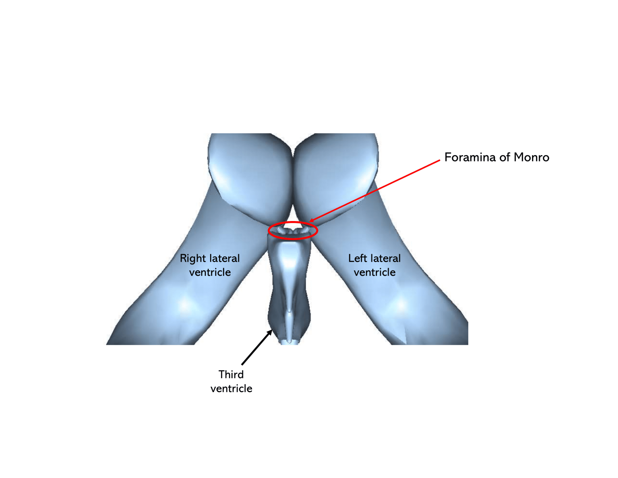
The interventricular foramen (or foramen of Monro) is a Y-shaped channel that connect the paired lateral ventricles with the third ventricle. The third ventricle is the narrow vertical cavity of the diencephalon. It is drained by the cerebral aqueduct (of Sylvius) that conveys CSF into the fourth ventricle.
Technology and Techniques in Radiology Foramen of monro in MRI
The foramen of Monro has also been referred to by the name of interventricular foramen. The structures comprising this foramen are the anterior part of the thalamus, the fornix and the choroid plexus.

Foramen The Definitive Guide Biology Dictionary
The interventricular foramen, also known as foramen of Monro , is part of the ventricular system and the connection between the third ventricle and the lateral ventricle . These paired foramina allow for the flow of cerebrospinal fluid between lateral ventricles and third ventricle s, and effacement or blockage results in non-communicating.
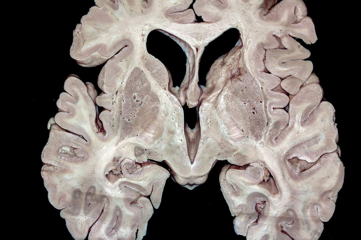
Coronal Section Across the Foramen of Monro and Apex of the Insula
The foramen of Monro has also been referred to by the name of interventricular foramen. The structures comprising this foramen are the anterior part of the thalamus, the fornix and the choroid plexus. Vital structures surround the foramen, the damage to which can be catastrophic leading to disability either temporary or permanent. In the literature it has been shown that tumors occurring in.
The Ventricular System Neuroanatomy CSF Geeky Medics
The differential diagnosis of masses arising from the foramen of Monro can be approached depending on the age of the patient. Pediatric choroid plexus papilloma adamantinomatous craniopharyngioma germinoma glioma Langerhans cell histiocytos.

Occlusion of one Monroi foramen leads to unilateral hydrocephalus
Introduction The foramen of Monro lies at the junction between the paired lateral ventricles and the third ventricle of the brain. Methods A comprehensive review of the literature was performed focusing on the foramen of Monro. Conclusions A good understanding of the anatomy of the foramen of Monro is essential for the neurosurgeon, especially with the increasing use of intraventricular endoscopy.

Illustration showing boundaries of the right foramen of Monro as
The interventricular foramen (aka Foramen of Monro) is a short passage extending from the lateral ventricle to the third ventricle, allowing for the drainage of cerebrospinal fluid from the lateral ventricles to the third ventricle. Complete Anatomy. The world's most advanced 3D anatomy platform.

Sectional Anatomy of the Brain Dr G R
The colloid cyst is a benign growth usually located in the third ventricle and at or near the foramen of Monroe, which is at the anterior aspect of the third ventricle of the brain. [1] The colloid cyst is an epithelial-lined cyst filled with gelatinous material. The gelatinous material commonly contains mucin, old blood, cholesterol, and ions.
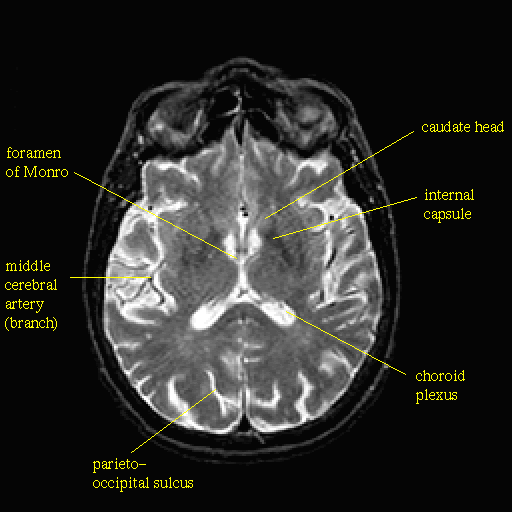
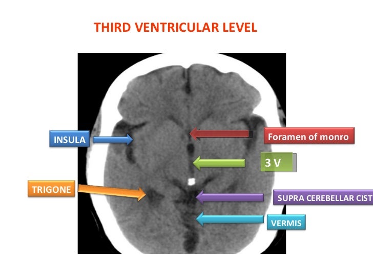
Axial Section of the Brain at the Level of the Foramina of Monro
The choroidal fissure forms a C-shape extending from the foramina of Monro to its inferior terminal point, which is termed the "inferior choroidal point." 9,15 Within the atria, there is a prominent triangular tuft called the glomus. 15 The tela choroidea is an invagination of the pia mater and ependyma, which gives rise to the choroid plexus within the choroidal fissure and along the roof.

Standard Endoscopic Vision of the Right Foramen of Monro Download
Findings: A sellar/suprasellar mass-like (long arrow) and rim calcifications (short arrows) impinging on the foramen of Monro causing hydrocephalus. (b): 20-year-old male with pineocytoma. Findings: A mass in the pineal gland (short arrows) with peripheral calcification (long arrow). (c): 13-year-old boy with pineal teratoma.
Ct basics
In the brain, the interventricular foramina (or foramina of Monro) are channels that connect the paired lateral ventricles with the third ventricle at the midline of the brain. As channels, they allow cerebrospinal fluid (CSF) produced in the lateral ventricles to reach the third ventricle and then the rest of the brain's ventricular system. The walls of the interventricular foramina also.

Anatomy of meninges, ventricles, cerebrospinal fluid
Introduction: The foramen of Monro lies at the junction between the paired lateral ventricles and the third ventricle of the brain. Methods: A comprehensive review of the literature was performed focusing on the foramen of Monro. Conclusions: A good understanding of the anatomy of the foramen of Monro is essential for the neurosurgeon, especially with the increasing use of intraventricular.